
Our lab has interdisciplinary biomedical engineering research programs focusing on the development of novel optical imaging methods for biomedical applications, including functional brain imaging, early cancer detection, cancer therapy monitoring and tissue engineering. Our optical imaging technologies include optical coherence tomography (OCT), multi-photon microscopy (MPM), fluorescence molecular imaging (FMI), fluorescence laminar optical tomography (FLOT), and endoscopy.
Technology Development
Early cancer detection
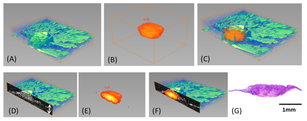
We integrated OCT with depth-resolved fluorescence laminar optical tomography (FLOT), which enables mesoscopic (mm-scale) morphological and molecular tomography. The hybrid OCT/FLOT imaging holds great promise as a powerful tool for the diagnosis of early cancers.
Cancer Therapy Monitoring
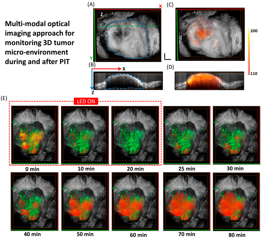
We applied a multi-modal optical imaging approach including high-resolution optical coherence tomography (OCT) and high-sensitivity fluorescence laminar optical tomography (FLOT), to provide 3D tumor micro-structure and micro-distribution of mAb-IR700 in the tumor simultaneously during photo-immunotherapy (PIT) in situ and in vivo.
Deep Brain Functional Imaging
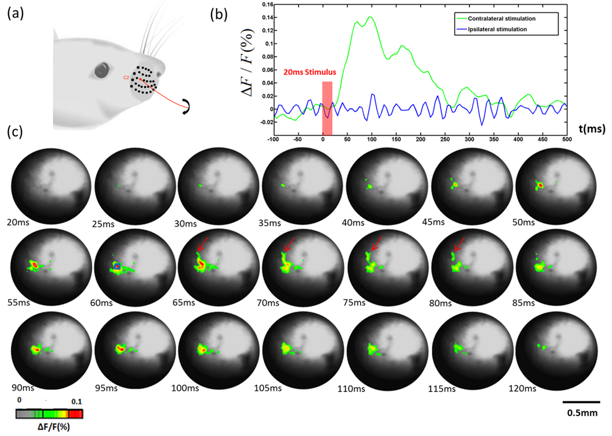
To access subcortical structures and image sensory-evoked neural activity, we designed a needle-based optical system using gradient-index (GRIN) rod lens. We performed voltage-sensitive dye imaging (VSDi) with GRIN rod lens to visualize neural activity evoked in the thalamic barreloids by deflection of whiskers in vivo.
3D Brain Functional Imaging in Mice Cortex
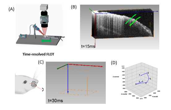
We applied angled FLOT (aFLOT) to record 3D neural activities evoked in the whisker system of mice by deflection of a single whisker in vivo. The results show that it is possible to obtain 3D functional maps of the sensory periphery in the brain. This approach can be broadly applicable to functional imaging of other brain structures.
FLOT in Bone Tissue Engineering
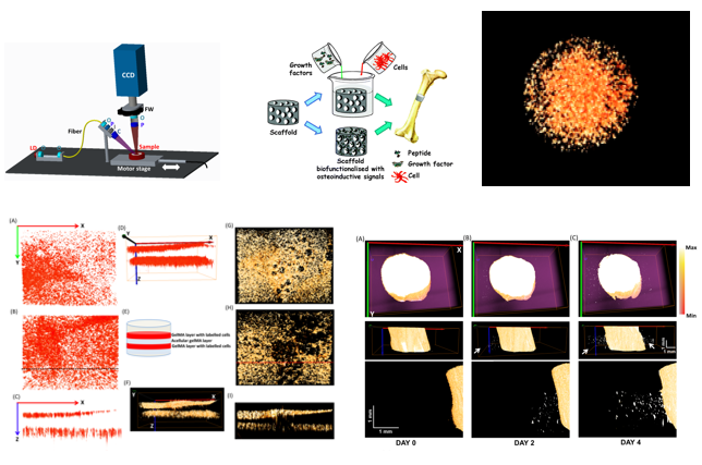
It remains a challenge to image three-dimensional (3-D) structures and functions of the cell-seeded scaffold in mesoscopic scale (>2 ∼ 3mm). We utilized angled fluorescence laminar optical tomography (aFLOT), which allows depth-resolved molecular characterization of engineered tissues in 3-D to investigate cell viability, migration, and bone mineralization within bone tissue engineering scaffolds in situ.
OCT Probe for Surgical Guidance
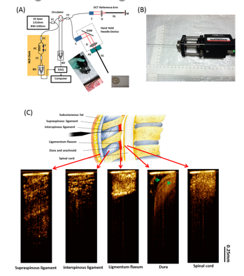
We developed a small hand-held optical coherence tomography (OCT) forward-imaging needle device for real-time epidural anesthesia surgery guidance and demonstrated its feasibility through exvivo and in vivo animal experiments. With tissue structures visualized and differentiated at the needle tip, OCT needle imaging device will enhance clinical outcomes with regards to complication rates, induced pain, and procedure failure when compared to standard practice.